
Cheap Dental Bone Graft Treatment in Sydney
There are several reasons that cause bone loss in the oral cavity. The affordable bone grafting procedure in Sydney is an excellent way to replace bone loss tissue and encourage natural bone growth. Affordable and Cheap Bone Grafting in Sydney is a versatile and predictable procedure nowadays.
Sometimes, bone graft is needed to create a stable base for dental implant placement, to halt the progression of gum disease, or to make the smile appear more aesthetically-pleasing.
Types of Cheap Bone Grafting Dental Treatment Offered by Us
Xenograft– This is the implantation of bovine (cow) bone. A Xenograft is safe and has been used successfully for many years. Ample bone can be obtained and no secondary donor site is necessary.
Allograft – Bone from a human tissue donor obtained from a bone bank (cadaver bone).
Autogenous bone graft – In this type of graft, the bone is removed from elsewhere in the body and implanted in the mouth. Common donor sites for bone grafting include the chin and wisdom tooth areas of the jaw. If large amounts of bone need to be harvested, the hip or the shin bone (tibia) is generally used.
Alloplast – Synthetic or artificial bone.
Reasons Why Affordable Bone Grafting May Be Necessary for Restoring the Jaw Bone
- Dental implants : Implants are the preferred replacement method for missing teeth because they restore full functionality to the mouth; however, implants need to be firmly anchored to the jawbone to be effective. If the jawbone lacks the necessary quality or quantity of bone, bone grafting can strengthen and thicken the implant site.
- Sinus lift graft : A sinus lift entails elevating the sinus membrane and grafting bone onto the sinus floor so that implants can be securely placed.
- Ridge augmentation : Ridges in the bone can occur due to trauma, injury, birth defects or severe periodontal disease. The bone graft is used to fill in the ridge and make the jawbone a uniform shape.
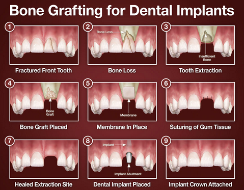
Bone Loss
Have you ever seen someone without their dentures in? Their face collapses inward. This is due to bone loss caused by loss of teeth. This process starts as soon as even one tooth is missing.
Over a short period of time, the jawbone associated with missing teeth dissolves and shrinks. Similar to a bar of soap in the shower, the bone “melts” away once a tooth is removed. 40-60% of the bone is lost in the first six months to one year after a tooth is extracted!
Shortly after the loss of teeth, the jawbone rapidly shrinks. Dental implants prevent and stop this continual bone loss. Even jaws like this can be rebuilt to have implants.
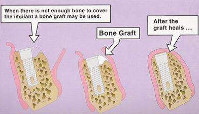
This often leaves a condition in which there is poor quality and quantity of bone suitable for placement of dental implants. In these situations, most patients are not candidates for placement of dental implants. Today, we have the ability to grow bone where needed. This not only gives us the opportunity to place implants of proper length and width, it also gives us a chance to restore functionality and esthetic appearance. If the bone is too thin or too short it must be replaced so an implant can be placed in the precise position of the tooth.
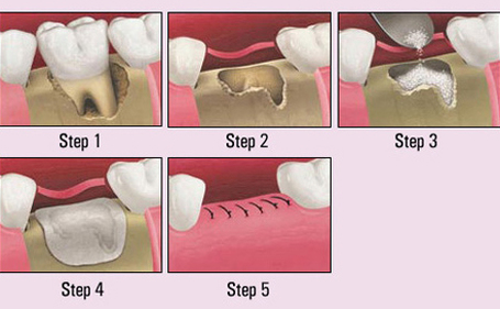
Extraction Socket Bone Graft
This is the most ideal way to prevent the inevitable bone loss that happens immediately after a tooth is removed. In this cost-effective, minimally invasive procedure, bone replacement is placed into the socket at the same time of the extraction. This is done if an implant cannot be placed at the same time.
Sometimes a “membrane” or covering is placed over the socket to prevent gums from growing inside the socket. Four to twelve weeks later, an implant can be placed in the exact position of the tooth.
This can also be done if a patient wants to have dental implants in the future and wants to slow the natural bone loss process.
Ridge Modification
Ridge modification is an effective procedure for treating bone loss in the upper and lower jaws. Bone loss occurs as a result of periodontal disease, trauma, injury, wearing dentures, or developmental problems. Such defects can leave insufficient bone for the placement of dental implants and an additional unattractive indentation in the jaw line adjacent to the missing teeth.
The bony defect can be filled with bone graft material which can help regenerate lost bone, or a bone substitute.
A few months of healing will be required for the bone to “take.” Depending on the case and type of implant and procedure, a dental implant may be placed during the ridge modification procedure or when healing is complete; much depends on the precise condition of the bone. Ridge modification improves the cosmetic appearance, functionality of the mouth, and the chance of enjoying dental implants for many years.
Sinus Lift Bone Graft
A type of bone replacement is called a Sinus Lift Bone Replacement Procedure.
Sinus Lift Bone Replacement Procedure:
Procedure 1. “Lateral Window or Traditional Approach” This common procedure and relatively painless procedure is only used to replace upper back teeth. The maxillary sinuses are behind your cheeks and on top of the upper teeth. Sinuses are like empty rooms that have nothing in them. Some of the roots of the natural upper teeth extend up into the maxillary sinuses. When these upper teeth are removed, there is often just a thin wall of bone separating the maxillary sinus and the mouth. Dental implants need bone to hold them in place. When the sinus wall is very thin, it is impossible to place dental implants in this bone.
There is a solution and it’s called a sinus bone graft or sinus lift. In this common procedure we enter the sinus from where the upper teeth used to be. The sinus membrane is then lifted upward and about a teaspoon of bone is inserted into the sinus. In many cases, the implants are placed at the same time. After several months of healing, the bone becomes part of the patient’s jaw and teeth can be placed on the implants.
The sinus graft makes it possible for many patients to have dental implants when years ago there was no other option other than wearing loose dentures.
If enough bone between the upper jaw ridge and the bottom of the sinus is available to stabilize the implant well, sinus augmentations and implant placement can often be performed as a single procedure. If the bone is very thin, the sinus bone graft will have to be performed first, and then the graft will have to mature for a few months, depending upon the type of graft material used. Once the graft has matured, the implants can be placed.
Procedure 2. Internal Sinus Lift ProcedureThis procedure is used when the bone loss of the upper back tooth area is minimal. In this minimally invasive procedure, the sinus bone is actually moved up through the implant site at the same time the implant is placed.
It is also possible to lift the sinus bone at the time of extraction of an upper molar in anticipation of a future implant. Bone replacement materials, if even necessary can be added through the tiny implant socket. Consult our office before you have an upper molar extracted as we can probably save you time and money.
Bone Widening/Expansion
In severe cases, the “ridge,” where the teeth were, has been lost and a bone graft is placed to increase ridge height and/or width. This is a technique used to restore the lost bone dimension when the jaw ridge gets too thin to place conventional implants. In this procedure, the bony ridge of the jaw is literally expanded by mechanical means.
This bone expansion is sometimes done as a separate procedure or it may be done at the same time the implants are placed. If it is done in two stages, usually only 3-4 weeks of bone healing is all that is necessary. The implants will be placed at either the first or second stage.
The bone graft may have to heal for a few weeks before attaching the teeth to the dental implants.
Bone grafting is a fairly simple procedure, which is usually done under sedation. Initially, the grafting material needs to either be harvested or prepared for insertion. An incision is made in the gum tissue. The bone grafting material is then placed at the affected site.
The bone regeneration process may be aided by:Gum/bone tissue regeneration – A thin barrier (collagen membrane) is placed below the gum line over the grafting material. This barrier creates enough space for healthy tissue to grow and separates the faster growing gum tissue from the slower growing fibers. This means that bone cells can migrate to the protected area and grow naturally.
Tissue stimulating proteins – Enamel matrix proteins occur during natural tooth development. Emdogain is a matrix protein product which may be placed on the affected site before the gum is sutured. It mediates the formation of accellular cementum on the tooth which provides a foundation to allow periodontal attachment to occur. Tissue stimulating proteins help to create lost support in areas affected by periodontal defects.
Platelet-rich growth factors – A high platelet concentration liquid can be used to create a blood clot at the site of a wound. It has recently been discovered that PRGF also stimulates bone growth – meaning a denser graft in a shorter time period.
The gum is sutured in place and a follow-up appointment will need to be made within 10 days to assess progress. Bone grafting is a highly successful treatment and a good base for further periodontal restorations.
If you have any questions about bone grafting, please ask us.
Now Let’s talk and review what a membrane is and its purpose.
A membrane is a barrier. Its purpose is to prevent gum from growing into the bone cavity. On many bone grafts for dental implants, a membrane is placed over the bone but under the gum.
There are many different types and styles of membranes but they break down into basically 2 categories.
- Dissolvable (resorbable) membranes
- Non-dissolvable (non-resorbable) membrane
Both resorbable and non-resorbable membranes have their use. A resorbable membrane will dissolve on its own. Non-resorbable membranes typically need to be extracted at some stage during your bone graft/dental implant procedure.
Resorbable membranes:
Most resorbable membranes used, as of 2014, are made of collagen. Collagen is a protein which you may have heard of in products such as shampoos and conditioners. It is heavily cross-linked and makes an excellent barrier. Most commercially available collagen membranes are 15 x 20mm in dimension and look like a flat, white piece of cardboard.
They are trimmed with surgical scissors by a dental implant specialist and typically covered over bone. Most collagen membranes are made from bovine Achilles tendon or porcine origin, which basically translates into cow and pig. Some resorbable membranes dissolve very quickly in just days; these are typically called plugs, while other resorbable membranes can last approximately 4 months. Most resorbable membranes are not tacked in place with titanium fixation tacks. Some are even pericardium from heart tissue.
There are also synthetic membranes that dissolve, such as Vicryl, which are not as commonly used.
Non-resorbable membranes:
Most non-resorbable membranes can be made of titanium and dPTFE (Dense polytetrafluoroethylene). They are materials that form a barrier that cannot be dissolved by your saliva. Some non-resorbable membranes, such as PTFE, are often combined with bone tacks in order to hold the membrane stable and cover the grafted bone.
One benefit of a non-dissolvable membrane– they are very predictable in generating bone. The main downside– it has to be removed at a second procedure. Sometimes that second procedure may involve just plucking out the membrane off the top of your socket graft or it may be more involved, such as completely reopening the gum, removing the bone tacks, and then removing the membrane. Typically when membrane removal involves removing tacks, the dental implants are placed at the same time that the membrane is removed.
PTFE has been used for over 30 years in cardiovascular applications such as suture, vascular grafts, and heart valves. PTFE is bioinert and does not cause inflammation.
Titanium reinforced membranes, or titanium mesh, is best at holding a space. It works like a tent and prevents collapsing of your gums to build new volume and mass underneath.
Sometimes the membrane on a socket graft will be removed after 1-4 weeks or may even fall out if it is a dPTFE style membrane. This is okay, generally. The purpose was to allow your gum to grow underneath it and cover over the bone graft as the membrane protects it. Once the membrane falls out or is removed from a socket bone graft, the bone still needs to heal for usually another 3-6 months before dental implant placement.
Is it okay that my membrane showing?
It depends. If the intention of your membrane was to be exposed from the beginning, then it is okay that it is exposed right now. If it was the intention for your gum to remain covering over the membrane for 4 -9 months, then you are likely having a minor complication called a dehiscence where the gum becomes exposed and naturally exposes the membrane and bone graft. The more critical the bone graft is, the more critical it is that the tissue remained closed and the membrane is not exposed at any time. Critical bone grafts would be onlay block bone graft, lateral window sinus bone grafts, nerve repositioning, and advanced guided bone regeneration (GBR).
A standard socket bone graft (where an outer wall is not missing) would not be considered a critical bone graft; so leaving a membrane exposed is okay. If an outer wall is missing, this would be considered a critical size defect.
It is important to maintain proper follow-up with your dentist throughout the procedure. It is likely that you will be on an antibiotic, such as amoxicillin or clindamycin, and a mouth rinse called chlorhexidine gluconate.
Keep in mind there are literally hundreds, even thousands of different membrane manufacturers each claiming to be better than one another. Yes, there are better manufacturers than others, but ultimately the surgical technique, training, judgment, and expertise of your dental implant specialist is going to be the most critical factor in the success of your bone graft…choose wisely!
You can use all of the best materials in the world, including growth factors such as PRP/PRF/PDGF/BMP, but in the hands of an inexperienced surgeon, results may not be that good. Of course your ability to heal is of paramount importance. Smoking never helps. Alcohol does not help either… Both of these slow down the healing of bone and greatly increase your chance of complications.
Wearing a removable, temporary “flipper” over a bone graft is to be done with extreme caution. Pressure from a removable temporary will cause the bone graft and membrane to move and almost always results in failure. Be sure you and your dentist speak about the use of a temporary and how to create space underneath it so there is no chance that it can apply pressure to the bone graft. Some bone graft membrane reconstructions are so critical and so large that the temporary cannot be worn at all, but that is the exception rather than the rule.
Tooth loss often leads to bone loss over time in the jaw. If the bone under the gum is not tall and/or wide enough to support dental implants, grafting will “build” enough bone for the implant procedure.
Your doctor will perform a complete medical history and physical examination before your surgery. Make sure you tell your doctor about any medications, over-the-counter drugs, or supplements you’re taking.
You’ll most likely be required to fast before surgery. This is done to prevent complications while you’re under anesthesia.
Your doctor will give you complete instructions about what to do the days before and the day of your surgery. It’s important to follow those instructions.
Your doctor will decide which type of bone graft will be used before your surgery. You’ll be given general anesthesia, which will put you into a deep, peaceful sleep. An anesthesiologist will monitor the anesthesia and your recovery.
Your surgeon will make an incision in the skin above where the graft is needed. Once the graft is securely in place, your surgeon will close the incision or wound with stitches and bandage the wound.
It usually takes six to nine months for an affordable bone grafting to fully heal and be ready for dental implants.
Affordable and cheap bone grafting in Sydney involves a small incision in the gum to gain access to the bone beneath it. Therefore, you may experience some post-operative soreness. Most people find this can be managed with ice packs applied to the jaw and over-the-counter anti-inflammatory medication and/or pain relievers.